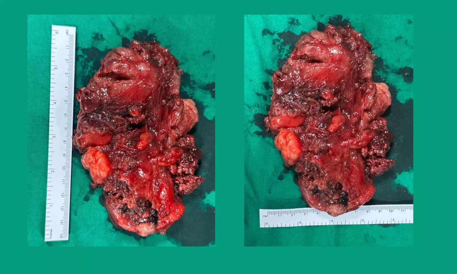
Rare 17 cm Ectopic Thyroid Tumour Removed from Chest Using Robotic Surgery at Sahyadri Hospital

Pune: In a rare and high-risk case, doctors at Sahyadri Super Speciality Hospital successfully removed a 17 x 12 x 11 cm, 800-gram ectopic tumour from the chest of a 62-year-old woman using advanced robotic-assisted surgery.
The tumour was diagnosed as an ectopic thyroid goitre, a condition where thyroid tissue develops in an abnormal location without any connection to the thyroid gland in the neck. This rare disorder is seen in only 1 out of 100,000 to 300,000 people.
The complex surgery was carried out by a multidisciplinary team at Sahyadri Hospitals, including Dr. Vinod Gore, Surgical Oncologist, Dr. Sanjay Kolte, Laparoscopic and Robotic Surgeon, Dr. Phulchand Pujari, Laparoscopic Surgeon, Dr. Swapnil Karne, Cardiothoracic Surgeon, Dr. Vikas Karne, Anaesthetist, and Dr. Balaji Momale, Senior Anaesthetist.
“The patient first came in with complaints of chest tightness. Scans showed a large tumour in the middle of the chest, in the area where the heart, windpipe, and major blood vessels are located.
What made the case truly exceptional was that her thyroid gland in the neck was completely normal, yet the tumour turned out to be thyroid tissue. Though it initially appeared to be a thymoma, it was ultimately identified in the thyroid biopsy as a rare ectopic thyroid goitre,” said Dr. Vinod Gore.
The tumour was situated near several critical structures, including the heart, superior vena cava, aortic arch, pulmonary arteries, and the pericardium. Due to its location, multiple hospitals had previously considered the tumour inoperable.
“To perform the surgery with high precision and minimal risk, we opted for a robotic-assisted approach,” Dr. Gore added. “This involved inserting robotic instruments through small cuts between the ribs, specifically in the 7th and 9th intercostal spaces.
The robotic system provided a 3D, magnified view of the chest cavity, offering superior control and flexibility. We had to rely entirely on visual cues and experience to avoid damaging nearby blood vessels, as robotic tools do not offer haptic (touch) feedback.”
Dr. Vikas Karne, HOD – Anaesthesiology, explained that airway management posed a major challenge. “The tumour had displaced the aortic arch and was compressing the trachea, making standard intubation extremely risky.
We performed an awake fibre-optic intubation, a highly specialised technique to secure the airway safely before administering anaesthesia.”
While most of the tumour was removed through robotic surgery, a portion near the top could not be accessed due to limited space and proximity to vital vessels. To complete the procedure, the team performed a mini-sternotomy, a small surgical incision in the upper breastbone, which allowed safe removal of the remaining tissue.
Dr. Sanjay Kolte noted, “The tumour was found to have no physical connection to the thyroid gland in the neck, confirming it as a true ectopic goitre, an extremely rare condition.
Its blood supply came from small vessels near the innominate vein and carotid arteries, which we carefully sealed during the procedure. One of the voice-controlling nerves, the left recurrent laryngeal nerve, was likely affected by pressure or stretching, while the right nerve remained intact.”
Dr. Phulchand Pujari highlighted the benefits of the approach: “The surgery was accomplished with minimal blood loss, and the patient did not require a transfusion or post-operative ventilator support. Pain management was effective, recovery was smooth, and the entire tumour was removed through a small chest incision. The sternum was closed using a single wire.”
Dr. Gore emphasised, “This surgery highlights the value of robotic technology in managing highly complex tumours. With careful planning, experienced hands, and the right tools, even rare and risky conditions like this can be treated effectively. Our goal was not just to remove the tumour, but to do it in the safest and most minimally invasive way possible.”
Dr. Swapnil Karne provided cardiac support throughout the operation, with a heart-lung machine on standby to ensure patient safety.
This case serves as an example of how advanced surgical techniques, thorough planning, and close teamwork across specialities can lead to successful outcomes even in highly challenging medical conditions.


