33-Year-Old Woman’s Brain Herniates Into Nose, Successfully Treated at HCMCT Manipal Hospital Dwarka

New Delhi: In a rare medical case, doctors at HCMCT Manipal Hospital Dwarka, Delhi, successfully treated a 33-year-old woman suffering from a severe condition in which a portion of her brain had herniated through the skull base into the nasal cavity and sinuses.
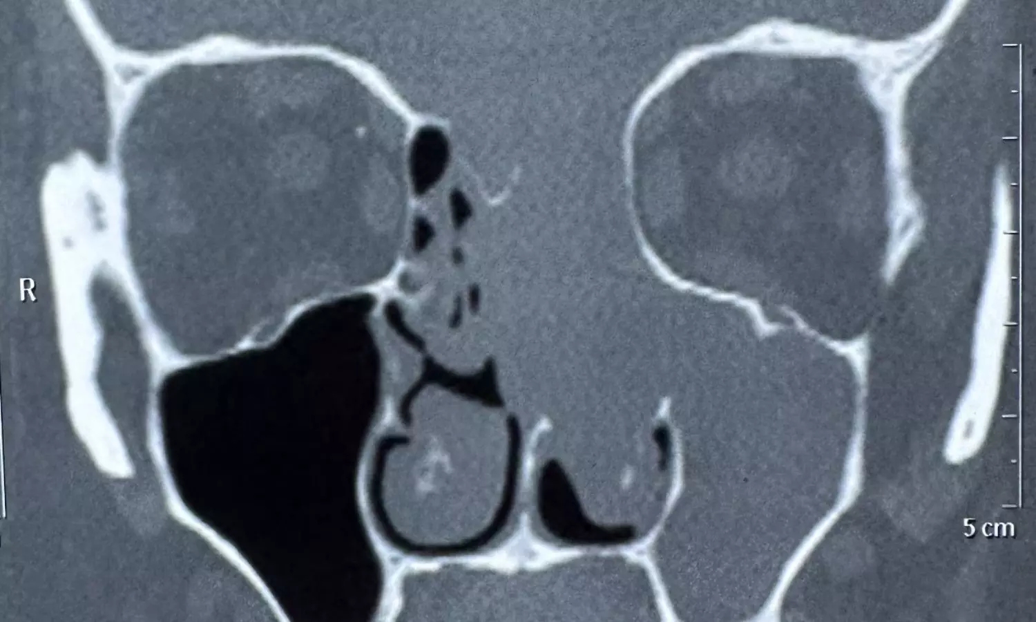
The brain tissue had descended into her nasal passage and oral cavity due to erosion of the cranial base. She came to the hospital with complaints of persistent nasal blockage and fluid discharge from the nose, which had been troubling her for several months.
The woman was suffering from nasal blockage caused by the gradual erosion of the cranial base (a thin bone separating the brain from the nasal cavity). This led to a rare and alarming situation where part of her brain slipped down and became visible through her nose and mouth.
The neurosurgery and ENT teams jointly performed an eight-hour surgery using advanced endoscopic and transcranial techniques to reposition the brain and rebuild the skull base. If this was left untreated, the condition could have caused fatal infections, meningitis, irreversible neurological damage, or severe bleeding.
Dr. Ashish Vashishth, HOD & Consultant - ENT (Otorhinolaryngology, Head and Neck and Cranial Base Surgery, Ear, Nose and Throat) said, “When the patient came to us with persistent nasal obstruction, an initial endoscopic examination in our ENT department raised suspicion of a rare condition.
Targeted CT and MRI scans revealed that part of her brain had herniated through the eroded skull base into the nasal cavity. This case certainly highlights the importance of a thorough diagnostic pathway and consulting the right specialists at the right time.
What made this case challenging was the technical precision needed to manage such a delicate and high-risk cranial base reconstruction. The success of this case truly highlights the strength of a multidisciplinary and diagnostic-first approach.”
Dr. Anurag Saxena, Cluster Head-Delhi NCR, Neurosurgery, said, “This was an extremely rare and technically demanding case. The patient was at significant risk of developing a life-threatening stroke, as the brain’s blood vessels were also herniating down along with the brain tissue.
Even the slightest injury to these vessels could have caused devastating complications. Almost half of the frontal and basal parts of the brain were hanging down through the skull base defect, which made performing the surgery exceptionally complex.
Through a carefully coordinated endoscopic and transcranial approach, we successfully repositioned the brain, reconstructed the skull base, and ensured her safe recovery. The patient has now recovered well and is leading a normal life under regular follow-up care.”


